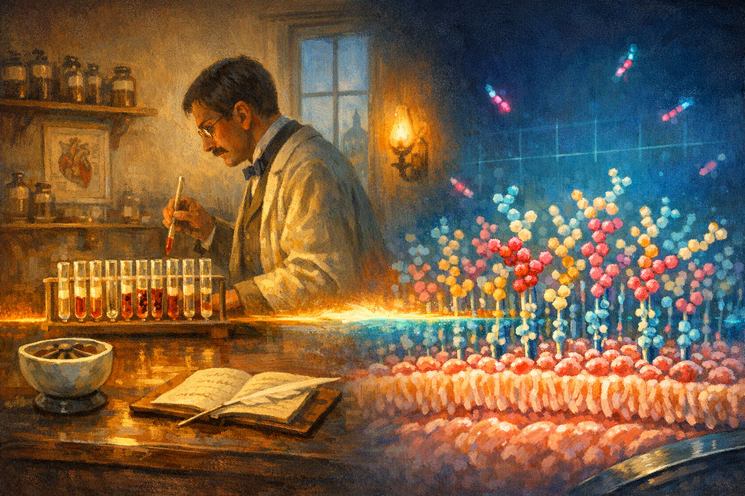
In the winter of 1900, in a small first-floor laboratory at the Institute of Pathological Anatomy on Spitalgasse in central Vienna, a thirty-two-year-old Austrian physician named Karl Landsteiner pricked the fingers of five of his colleagues, separated the painterly red cells from the serum in each sample with a small porcelain centrifuge, and began to mix every serum with every set of cells in a grid of small glass test tubes. He was looking for the reason that blood transfusions had been killing roughly half of the people who received them.
Some tubes did nothing. The serum and the red cells sat together quietly. The tubes that did something turned cloudy. The red cells in those tubes had clumped into small dark grains visible against the cream-coloured serum, as if the cells had been recognised as foreign and condemned by the chemistry of the donor. Landsteiner recognised at once that he had a sorting principle.
He wrote up the result the following autumn as a footnote in a longer paper on the agglutinating action of normal blood serum, published in the German journal Zentralblatt für Bakteriologie in 1900. The full account followed in 1901 in the Wiener klinische Wochenschrift under the title Ueber Agglutinationserscheinungen normalen menschlichen Blutes, on the agglutinating phenomena of normal human blood. He had identified three groups, which he called A, B and C. The C group was relabelled O within a few years, because the clumping pattern made it the absence of either of the other two antigens. A fourth group, AB, the rarest, was identified the following year by two of his colleagues, Alfred von Decastello and Adriano Sturli, working in the same Vienna laboratory.
The reason for the clumping was a sugar. Not the sugar you can taste. A sugar molecule attached to the outside of the red blood cell, sitting at the very tip of a slim chain of other sugars that protruded into the bloodstream. Group A had one sugar at the tip. Group B had another. Group O had neither. Group AB had both. The painterly chemistry the immune system was reading, and either tolerating or rejecting, was sugar chemistry on the surfaces of cells.
The medical world essentially ignored the result. Surgeons continued to transfuse blood without typing it, and patients continued to die at roughly the same rate, until 1907, when an obstetrician at Mount Sinai Hospital in New York named Reuben Ottenberg performed the first deliberately matched transfusion, having read Landsteiner's paper. Ottenberg used the technique on hundreds of patients over the next several years and almost never lost one. By the First World War the British and French and Americans were typing blood at field hospitals; by the second, the typing was being done in the back of a moving ambulance. Tens of millions of lives have now been saved by Landsteiner's footnote. He was awarded the Nobel Prize in Physiology or Medicine in 1930.
The discovery did something quieter than save lives. It announced that the outside of a cell was not a featureless membrane. The cell was wearing a fingerprint.
The fingerprint is made of sugars. Not the kind that sweeten coffee. They are short chains of saccharide molecules called glycans, anywhere from one to a few dozen units long, attached to the proteins and the lipids of the cell membrane. Together they form a dense forest called the glycocalyx, three to five hundred nanometres thick, that every animal cell wears on its outer surface. Every cell type in the human body wears a slightly different glycocalyx, and the glycocalyx of any given cell changes as the cell's situation changes. Cancer cells re-pattern their glycans as they become malignant. Immune cells re-pattern theirs when they are activated. Stem cells re-pattern theirs as they differentiate. The information was always there. We could not read it.
This month, in Nature Nanotechnology, a team led by Leonhard Möckl at the Max Planck Institute for the Science of Light in Erlangen has reported the first method that can. It is called Glycan Atlasing. The technique stitches together two ideas. First, the team designs DNA-tagged lectins, sugar-binding proteins from plants and bacteria each of which recognises a specific glycan structure, and uses them as labels. Second, they pair the labels with metabolic oligosaccharide engineering, a technique that feeds the living cell sugars with small chemical handles attached, so that the cell incorporates the handles into its own glycocalyx as it grows. Together, the two techniques let the researchers see, under a super-resolution microscope, the actual position of every recognisable sugar on the surface of a cell at a resolution of a few nanometres.
The result is an atlas. They have mapped the glycocalyx of dozens of cell types from across the human body and shown that the spatial pattern of sugars, not just which sugars are there, reliably tracks what the cell is doing. Cancer cells from a breast tumour rearrange their surface sugars in ways that distinguish them clearly from the surrounding healthy tissue, even before the cells have started to multiply detectably. Activated immune cells re-pattern their surface within hours of seeing a pathogen. The Möckl group is now working with two German university hospitals on whether a routine blood sample, run through the Glycan Atlasing pipeline, could detect early-stage cancers that would otherwise not be visible for years.
If the technique scales, the medical implications are quietly enormous. Cancer detected from a blood sample is cancer that surgery can still cure. The Möckl paper is not yet that test; it is the proof that the test is possible.
What it adds to the longer story is what Landsteiner added in 1900. The sugars on the surfaces of cells are how cells talk about themselves. The body has been carrying a chemical fingerprint, on every cell of every tissue, since before there were doctors to ask about it. Landsteiner found four patterns and saved tens of millions of lives. Möckl's team has now mapped thousands. The patterns were there the whole time.
There is a quiet sentence at the end of Möckl's paper that is worth quoting. The authors write that the glycocalyx, even with the new instruments, is still a long way from being completely characterised. They write that they have produced an atlas, not a map. There is a difference. An atlas shows where the major regions are; a map shows what is in them. Landsteiner produced an atlas in 1900. The next twenty years filled it in. The same will be true of this one.
The sugars were doing the talking the whole time. We are only now beginning to write down what they said.