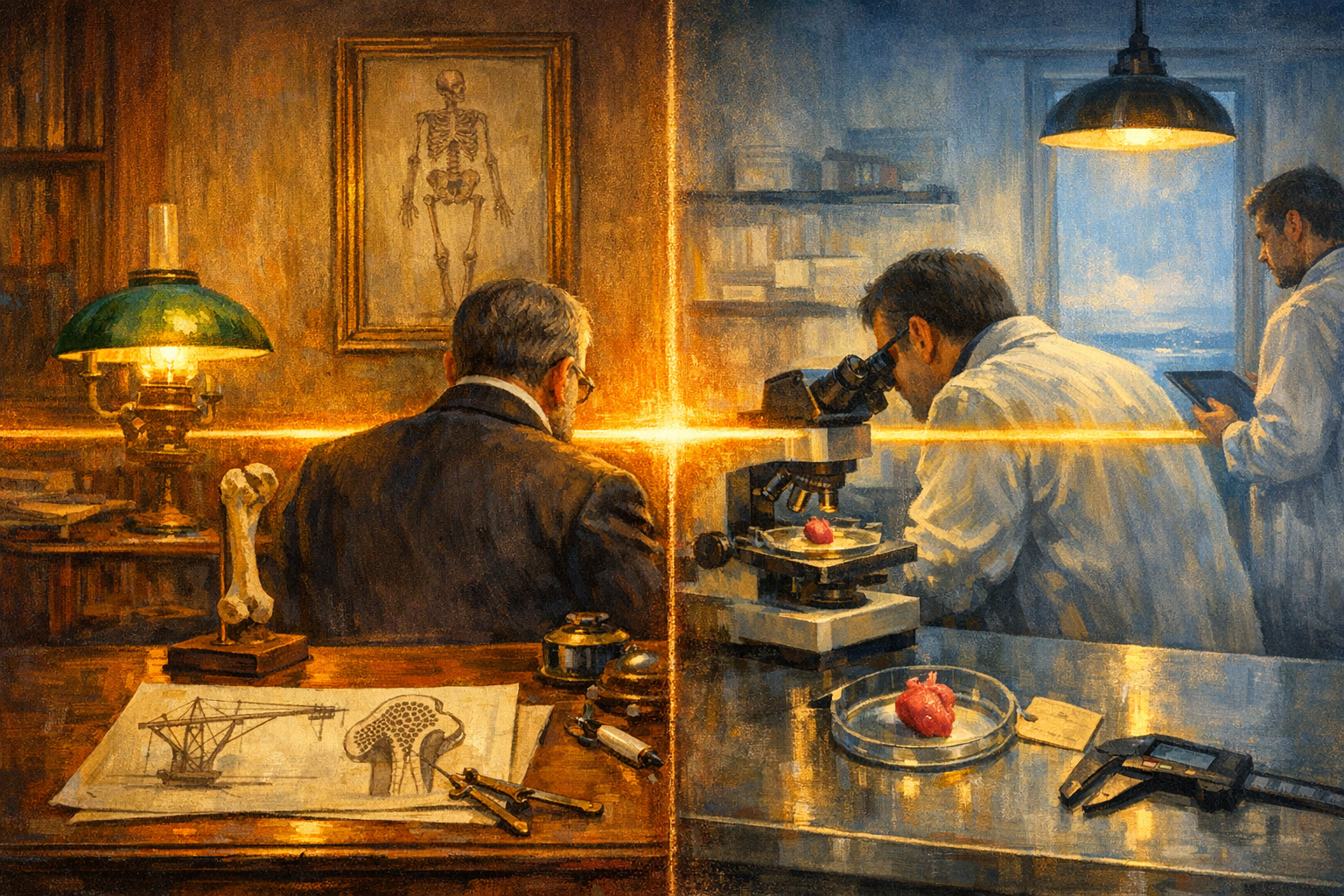
In the spring of 1866, a Bavarian engineer named Carl Culmann, who held the chair of structural mechanics at the new Polytechnikum in Zurich, finished a book called Die graphische Statik. It was the first book on graphic statics, a method of drawing the forces inside a loaded beam or truss as visible lines. Culmann's textbook contained an idealised drawing of a Fairbairn crane, a curved cast-iron jib of the kind then used in dockyards from Newcastle to Trieste, with the stress trajectories inked through the body of the metal. The lines arched up from the foot of the jib, crossed at right angles, and converged on the point where the load hung. A crane under load looks, on paper, like a piece of latticework.
The following year, an anatomist at the University of Zurich named Georg Hermann von Meyer published a short paper in which he placed Culmann's crane diagram next to a coronal cross section of the human femur. The two pictures were almost the same picture. The trabeculae inside the head and neck of the thigh bone, the bony lattice that holds up the femoral head where it socket-joints into the pelvis, arched in the same direction as Culmann's stress trajectories, crossed each other at the same right angles, and converged on the same load point. Von Meyer had spent his career staring at femoral cross sections without knowing what he was looking at. After one conversation with Culmann at a dinner in Zurich, he knew.
Twenty-five years passed before anyone in medicine took the analogy seriously. The man who did was Julius Wolff, professor of surgery at the Friedrich Wilhelm University in Berlin and the director of the new orthopaedic clinic that would in 1902 be folded into the Charité. Wolff was a working surgeon. He had set broken legs in the Franco-Prussian War. He had watched bones heal in patterns that did not match the textbooks. In 1892, in a monograph called Das Gesetz der Transformation der Knochen, he proposed something his colleagues found difficult to accept. Bone, he wrote, is not a static structure. Bone is a working material that actively remodels itself to match the mechanical loads applied to it. Load a femur and the trabeculae thicken along the lines of force. Stop loading it and the trabeculae thin out. The Culmann crane and the von Meyer femur were not a coincidence. They were the same thing.
The proposition was treated as a pretty analogy for the next eighty years. The mechanism was nowhere. Cells were thought to read chemicals, not forces. By what means could the inside of a thigh bone know which way was up?
It took until the 1990s for that question to get a molecular answer. The discovery of integrins and focal adhesions in the 1980s showed cells anchoring themselves to neighbouring tissue through specific proteins. The Piezo ion channels, found in 2010 in the Scripps laboratory of Ardem Patapoutian, turned out to be specifically responsive to physical deformation of the cell membrane. Patapoutian shared the 2021 Nobel prize in physiology for that discovery. By the time Patapoutian was on the podium in Stockholm, mechanobiology had become its own field, with its own journals and its own conferences. Wolff's law had a molecular basis. Cells genuinely do read force.
Last month it became clear that they read it in a much more consequential place than anyone had supposed.
On 24 April 2026, a team led by Giulio Ciucci at the International Centre for Genetic Engineering and Biotechnology in Trieste published in Science an answer to a question oncologists had circled for a hundred years. Primary cancers of the heart are extraordinarily rare. Autopsy series put the incidence at roughly two in ten thousand. The heart is a large, well-vascularised, hard-working organ. By every other measure it ought to develop tumours about as often as the kidney or the liver. It does not. The standard guess, in textbook after textbook, was that cardiomyocytes mostly stop dividing in adulthood, so there are few opportunities for the mutations that cause cancer to take hold. The new paper says the standard guess is at most half the story. The other half is the beat itself.
Ciucci's team built a small experimental system that separated the heart's two roles. They transplanted a donor mouse heart into the neck of another mouse, where it received blood supply and stayed alive but did not mechanically pump. Then they injected human lung cancer cells into both the recipient mouse's original beating heart and into the transplanted non-beating heart. In the heart that was not pumping, the cancer cells ran riot. In the heart that was, they replaced only about a fifth of the tissue and then stopped. The difference was not chemistry. The blood supply was the same. The difference was the squeeze.
When the researchers looked for the molecule that does the squeeze-detection, they found Nesprin-2. Nesprin-2 sits in the outer membrane of the nucleus. It is the heart of a small assembly called the LINC complex, which acts as a mechanical bridge between the protein skeleton of the cell and the protein scaffolding of the nucleus itself. Every time the heart contracts, the entire cardiomyocyte is briefly compressed, the cytoskeleton tugs on the LINC complex, and Nesprin-2 transmits the pulse into the nucleus. The chromatin inside, the DNA wound around its histone proteins, becomes less compact. Genes that would normally stay locked away, including a set of genes that brake cell division, become accessible and get expressed. The cancer cells inside the beating heart are not killed. They are told to stop. When Ciucci's team silenced Nesprin-2 in the beating heart, the brake disappeared and the cancer took off.
The implication is straightforward enough that it makes oncologists nervous to say it out loud. Mechanical force, applied at the right frequency, can suppress cancer-cell proliferation in tissue that does not normally experience that kind of force. Several drug-development paths are now open. One is to find a small molecule that mimics the chromatin-loosening effect of Nesprin-2 tension. Another is to find a device that applies physical pressure pulses to soft-tissue tumours in vivo. Neither would be a cure, and oncology has had its fingers burned on simple mechanisms before, but the proposition that you might one day prescribe a heartbeat to a tumour is no longer absurd.
It is also no longer original. Julius Wolff drew the conclusion in Berlin in 1892, from a comparison of human thigh bones with a dockyard crane drawn in Zurich. Living tissue, he proposed, listens to force. We took 130 years to find the protein that does the listening. We have just discovered that the same protein, in a different organ, is the reason the heart almost never gets cancer.
The trabeculae inside your femur run along the lines of force. So do the genes inside your heart.